It is a closed bone box inside the inner ear that protects the hearing and stability organ. The disease of this structure, which progresses with the loss of bone tissue and new unsystematic bone formation due to genetic or environmental factors, is called otosclerosis.
Sound waves pass through the external ear canal and vibrate the membrane. The eardrum separates the outer ear and middle ear cavity from each other like a tight curtain. The middle ear cavity is filled with air, and the hammer, anvil, and stirrup are responsible for transmitting sound vibration between the eardrum and the middle of the inner ear. The stirrup closes an opening to the inner ear called the oval window. From this opening, sound waves pass into the fluid-filled inner ear hearing organ.
Otosclerosis disease; This causes new unsystematic bone formation around the oval window and the stirrup, reducing the transmission of sound to the inner ear.
This disease is more common especially in young women aged 15-45 years. Apart from genetic features, measles infection and hormonal changes (such as pregnancy) cause the disease to progress and cause complaints.
The most common symptom is progressive hearing loss, which we call conductive type. It is typical for hearing to be more adequate in crowded loud environments than in quiet environments. Apart from hearing loss, some patients may also complain of imbalance. In approximately 70% of patients, otosclerosis can present in both ears. The degree of hearing loss usually comes to a certain level and stabilizes. In very few patient groups, progressive otosclerosis envelops the inner ear auditory organ and causes total hearing loss in that ear.
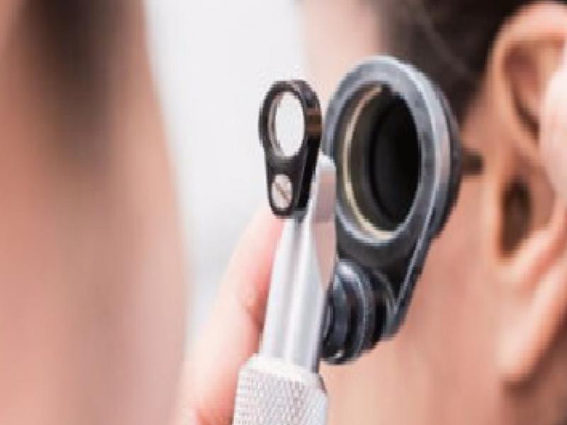
ENT examination, hearing tests and radiological imaging methods are used for diagnosis. It is typical that the eardrum is normal on examination, and sometimes a red reflex is seen behind the eardrum. The degree and type of hearing loss should be determined by audiological hearing test. Typical low ear pressure in ear pressure tests and negative acoustic reflex test are characteristic features. Computed ear tomography is a valuable diagnostic test to distinguish the other causes of hearing loss behind the intact eardrum and to detect the otosclerosis focus.
A complete curative procedure to eliminate otosclerosis has still not been found. Treatment is planned to correct the hearing loss caused by it.
Types of treatment
-
Follow-up
-
Medication
-
Hearing aid
-
Surgical
Follow-up is an option, especially in patients with new onset and very mild hearing loss. This disease is a disease of the inner ear bone and occurs as a result of bone destruction and metabolic changes in the manufacturing face. Drug treatments used in bone resorption such as osteoporosis; It helps to stop the progression of hearing loss. However, it has no curative properties. Long-term use of sodium fluoride and bisphosphonate derivatives is required.
When the goal is to restore hearing, there are two alternatives: Hearing aid and surgery.
With the operation, the intact eardrum is opened and the middle ear cavity is entered. In this way, other diseases that can cause hearing loss in the middle ear can be diagnosed. It is diagnostically valuable to see that the stirrup that is seen in otosclerosis does not vibrate and is fixed. In this case, some or all of the stirrups are removed. A new hole is made in the fixed oval window and the inner ear is entered. Afterwards, the Teflon piston prosthesis suitable for the patient’s anatomy is placed in the middle of the anvil bone through this hole. Thus, the sound wave vibration is transmitted directly from the anvil to the inner ear with the help of the prosthesis.

