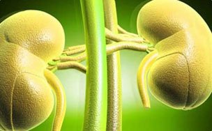
Symptoms in kidney diseases can be classified as changes in micturition habits, changes in urine amount, changes in urine composition, symptoms resulting from kidney dysfunction, pain and edema.
Micturation disorders (voiding disorders)
The most common complaint related to the habit of urination is frequent urination and is defined as “frequency”. This situation can be seen with increased urine amount (polyuria) or in the presence of normal urine amount. Frequent urination in the presence of normal urine volume may be the result of inflammation, irritation of the bladder as a result of stones or tumors, decreased bladder capacity due to fibrotic contraction (fibrosis after radiotherapy), external pressure on the bladder (pelvic mass, gravid uterus). If the complaint of frequent urination is accompanied by a large amount of urine each time, it brings to mind the association of polyuria, if it is accompanied by less urine, a bladder capacity disorder. Frequent urination is often associated with nocturnal micturition. Nocturia can also be seen in patients with sleep disorders. Normally, sleep stimulates the release of ADH, so the amount of urine decreases during sleep. ADH secretion does not increase in patients who cannot sleep even though they lie down to lie down, renal blood flow increases in the recumbent position, and the patient has to urinate at night as a result of increased urine production.
Prostatic enlargement in middle-aged men may cause complaints such as weakening of the urine flow, difficulty in starting to urinate (hesitancy), continuation of the dripping flow when the urine is finished (terminal dribbling), bifurcation of the urine. If the urethral obstruction becomes complete, it may result in urinary retention, acute bladder distension, and bilateral hydronephrosis. On the other hand, in some cases with prostatic hypertrophy that has caused incomplete obstruction, urinary retention and retrograde pressure reflection, and accordingly, a decrease in the filtrate flow of the nephrons, a deterioration in the composition of the medulla that helps to maintain its concentration ability, and as a result, an increase in the amount of urine occurs. Paradoxically, this increase in the amount of urine, which can be seen in cases with incomplete obstruction, can cause a delay in diagnosis, often as a result of wrong evaluation.
The presence of pain and discomfort during the urination process is referred to as inflammatory urine, painful urine (dysuria). Cases often describe it as a burning or stinging sensation in the urethral meatus or suprapubic region during or just after voiding. This complaint usually develops as a result of bladder, prostate or urethral inflammation. If the complaint of urinary inflamed (dysuria) is accompanied by complaints such as frequent urination, feeling the need to urinate immediately (frequncy, urgency), it indicates the presence of cystitis. This condition is most common in young women and often associated with sexual activity. Structural disorders and diseases related to the bladder or prostate are usually found under such complaints in elderly women or men. In men, perineal or rectal pain should suggest the presence of prostate inflammation. In the presence of inflammation, crying, or an unexplained fever during voiding in children and young adults, urinary tract infection should be suspected and structural anomalies of the urinary tract should be sought.
Urine quantity disorders
An increase in daily urine amount is defined as polyuria (polyuria), a decrease as oliguria (oliguria), and its absence as anuria (anuria). The average daily urine amount is related to environmental factors and daily water intake habits under normal conditions. In a study conducted by Istanbul Medical Faculty, Department of Nephrology, the average daily urine amount of 32 normal adults was found to be around 1200 ml. A daily urine output of more than 2 liters is called polyuria. Normal people can become polyuric when they drink too much water, tea, coffee, and alcohol. As other important causes of polyuria;
Excessive drinking of water as seen in the example of primary polydipsia
Increased tubular solute load (urea in the presence of chronic renal failure, glucose in the presence of hyperglycemia, low molecular weight proteins in myeloma)
A decrease in ADH production decrease (head trauma, tumors, hypothalamus or pituitary infections)/Central Diabetes Insipidus
Disruption of medullary concentration gradient as a result of renal medullary disease (nephrocalcinosis, analgesic nephropathy, renal papillary necrosis, medullary cystic disease, sickle cell disease, urinary tract disease) incomplete obstruction)
Impairment of tubular cell response to ADH (hypercalcemia, hypokalemia, lithium toxicity, ifosfamide toxicity, congenital nephrogenic diabetes insipidus, Sjögren’s syndrome, Cushing’s syndrome)
Polyuric healing phase of acute tubular necrosis
Bed rest and diuretic use during edema treatment
Mannitol and hypertonic glucose infusions
can be counted. The production and removal of urine below the amount of urine required for the elimination of the solute load produced as a result of the daily normal metabolic function constitutes the theoretical/physiopathological definition of oliguria. The amount of urine output below 400 ml and above 50 ml per day in adults is accepted as oliguria, and this indicates the presence of acute renal failure due to prerenal causes, acute vasculitis, acute glomerular lesions, tubular necrosis caused by toxin or sepsis, acute interstitial nephritis.
Daily urine being less than 50 ml and anuria (no urination is called complete anuria) firstly bring to mind shock and bilateral urinary tract obstruction (prostate, pelvic tumors). Rarely, anuria may be seen in the presence of renal infarction/cortical necrosis, severe acute vasculitis, Good pasture syndrome, and hemolytic uremic syndrome. Older chronic kidney patients receiving renal replacement therapy such as hemodialysis also often have oliguria or anuria.
Changes in urine composition
Classically, the presence of >3 erythrocytes in men and >5 erythrocytes in women in each field during IM examination of the urine sediment is defined as hematuria, but nowadays, the presence of more than 2 erythrocytes in each augmentation field with IM It is emphasized as abnormal for both sexes. The presence of visible hematuria is one of the important complaints that promptly takes the cases to the physician, and it may originate at any level from the glomeruli to the urethral meatus. Infection, stones, tumors and glomerulopathies are the main causes of hematuria. Hematuria may also be seen in conditions such as bleeding diathesis, use of anticoagulants, vascular anomalies and sickle cell disease. While hematuria is detected only by microscopic examination in some cases, it can be noticed with the naked eye in some cases (macroscopic hematuria). Macroscopic or gross hematuria does not mean significant blood loss. A small amount of blood as small as 1 ml can change the color of 1 liter of urine perceptibly. Hematuria originating from the glomeruli often causes the urine to become red-brown in color, sometimes the color change due to glomerular hematuria can be defined as turbid, tea or coca-cola color. Macroscopic hematuria is intermittent rather than continuous. This condition often develops in cases of IgA nephropathy and especially during a mucosal infection such as an acute upper respiratory tract infection and usually resolves within 1-3 days. In the period between attacks, microscopic hematuria usually persists. It should be remembered that hematuria arising from urethral pathologies manifests itself immediately at the beginning of urination, and if the urine is collected as first stream, mid stream and last stream sample (three goblet test), the probability of detecting hematuria (initial hematuria) in the first urine sample is higher. Hematuria due to bladder and prostate pathologies is more often noticed towards the end of the micturation process and this feature is referred to as terminal hematuria. Detection of blood clots in the urine is an important finding in eliminating hematuria of glomerular origin, and its presence should first bring to mind bladder tumors. In marathoners and serious joggers, transient hematuria can be identified, possibly due to mild irritation of the bladder mucosa. While some cases are being investigated with the preliminary diagnosis of hematuria, it is noticed that they have hemospermia. This finding usually develops as a result of prostate pathology or bleeding diathesis. Red-brown urine color change does not always develop due to hematuria, but may also be seen for other reasons (Table-1). The differential diagnosis of this will be made later.
Table-1: Main causes of red-brown color change in urine color
Hematuria
Hemoglobinuria
Myoglobinuria
Urates
Porphyria
Alkaptonuria (homogenetic acid)
Medications (phenacetin, antipyrine, rifampicin, metronidazole, nitrofurantoin, warfarin, phenytoin)
, some vegetables
, various food dyes)
The presence of proteinuria in the urine is usually determined chemically, but some patients notice that their urine is foamy. Normally, up to 150 mg of protein can be found in 24-hour urine, and more than 50% of this is of tubular origin. Investigation of the presence of protein in the urine in terms of screening is commonly done by the “stix” test all over the world. Since albuminuria is determined with this test, it should be remembered that this test will be negative in myeloma cases, whereas the precipitation tests will be positive. It is known that in some normal cases, mild (< 1 g/day) proteinuria may occur without any pathological significance. In some cases, proteinuria was related to position and it was shown that proteinuria was not found in the urine collected during bed rest, but proteinuria was determined in the urine collected during daily activity. This type of proteinuria is defined as orthostatic or postural proteinuria, and this condition is reported to have a benign course. It is claimed that the “nutcracker” phenomenon plays a role in some orthostatic proteinuria cases. Another benign type of proteinuria is the presence of proteinuria that develops only after exercise. Pathological proteinuria indicates a glomerular or interstitial disease. Proteinuria that develops as a result of interstitial disease is usually mild and is less than 2 g per day. It is very rare to encounter nephrotic level proteinuria due to interstitial disease. In multiple myeloma, severe proteinuria can be seen due to renal involvement in the form of cast nephropathy, which is referred to as pseudonephrotic syndrome. In proteinuria of glomerular origin, the amount is variable and may be at serious levels (such as 10 g and above). The type of proteinuria can sometimes be diagnostic. For example; Presence of selective proteinuria suggests minimally altered disease, while Bence-Jones proteinuria indicates myeloma.
Bacteriuria (presence of bacteria in the urine); may be symptomatic or asymptomatic. Urine collected in the bladder is normally sterile. The urethra, and especially the urethral meatus, is not sterile and urine may be contaminated during micturation. Therefore, when the case is asymptomatic, bacteriuria is detected in ml of mid-stream urine sample taken from the first urine in the morning in a sterile container. It is defined as significant bacteriuria when there is more than 100000 (105) bacterial growth in
Leukocyturia; It is a definition used when more than 3-5 leukocytes are seen in each light microscopy field in the examination of urine sediment. Besides urinary system infection, it is a nonspecific finding that can be seen in many diseases such as nephrocalcinosis, papillary necrosis, analgesic nephropathy, polycystic kidney disease, interstitial nephritis. Severe leukocyturia is defined as pyuria. The presence of pyuria more strongly indicates infection. The presence of sterile pyuria can be seen in tuberculosis and chlamydial infections. The main causes of sterile pyuria are given in Table-2.
Stones can be expelled spontaneously with urine. This condition is usually accompanied by colic-like pain, but sometimes it can be painless. Small tissue particles may also come with urine, as can be seen in papillary necrosis and urinary tract tumors.
Pain
Pain is not a very valid complaint in terms of showing all kidney diseases, and inflammation and obstruction should be considered most often in its presence. In the form of pyelonephritis, kidney inflammation usually causes localized pain in the area designated as the kidney angle on the affected side. This pain develops gradually, although the degree of severity is variable, it is usually of a constant character. In the presence of perirenal abscess, if the abscess develops upwards and irritates the diaphragm, its symptoms appear. If it develops downwards, irritation findings of the psoas muscle are observed. Glomerular inflammation usually does not cause pain but may be accompanied by a blunt flank pain, particularly in the case of acute glomerulonephritis and IgA nephropathy.
Some cases occasionally complain of a blunt flank pain of varying severity, accompanied by a noticeable hematuria. This condition is defined as “loin pain hematuria” syndrome. C3 accumulation in the afferent arteriole wall and some nonspecific findings can be seen in kidney biopsy. The meaning and significance of C3 deposition in the afferent arteriole wall has not yet been clarified. In such cases, renal angiography may show bent, stenotic or occlusive lesions. There are authors who claim that the presence of chronic, persistent pain, especially in the left flank, without hematuria, may be associated with the “nutcracker phenomenon”.
The pain that develops in acute ureteral obstruction is usually sudden, severe and colic and spreads to the groin and scrotum. In chronic obstructions, there is usually no pain. The classic signs in the presence of urolithiasis are renal colic and hematuria. In addition to these symptoms, there may be complaints such as abdominal pain, nausea, difficulty urinating, penile pain or testicular pain.
In most kidney diseases, no complaints such as pain or discomfort in the flanks are encountered. Without pain, kidney functions may be severely impaired and cases may present with metabolic consequences of severely impaired kidney functions.
Edema
Edema can be defined as a palpable swelling that develops as a result of increased interstitial fluid volume. The main renal syndromes that may be associated with edema are known as nephrotic syndrome, acute renal failure, chronic renal failure and acute nephritic syndrome. In addition to these renal causes, congestive heart failure and cirrhosis are among the other important causes of edema. While edema usually becomes prominent around the eyes in the morning, it develops in the legs and ankles later in the day. This diurnal change disappears as the amount of edema increases. Peripheral edema examination is performed from the pretibial surfaces of both lower extremities and from the sacrum in continuously lying cases.
Symptoms of hypertension
Renal parenchymal diseases of glomerular origin are usually associated with hypertension. Hypertension is less common in renal diseases of tubular and interstitial pathologies. Hypertension is often found in the late stages of chronic renal failure. Hypertension may reflect itself with complaints such as headache, dizziness, ringing in the ears. Sometimes, it may present with signs and symptoms of acute left heart failure such as shortness of breath and palpitations. Severe high blood pressure may be present as a sign of a systemic disease such as systemic sclerosis. Presence of renal artery stenosis should definitely be remembered in patients who present to the emergency room with recurrent pulmonary edema and no serious cardiac pathology can be demonstrated.
Uremic manifestations
Some cases present with uremia as a result of impaired renal function. Uremic signs and symptoms; anorexia, tiredness, weakness, nausea, vomiting, hiccups, taste disorder, itching, irritability, insomnia, dirty-pale skin color, pale mucous membranes, dry skin, traces of itching on the skin, hypertension, pleural or precordial smearing, ammonia breath, flapping tremor, confusion, changes in consciousness and personality up to coma, acidotic respiration, shortness of breath, palpitation, epistaxis, melena, hematemesis, delayed first menstruation, amenorrhea, menorrhagia, infertility, restless legs, loss of libido, impotence.
Presence of renal involvement accompanying systemic diseases
Many systemic diseases cause renal involvement during their emergence or course. In addition to the signs and symptoms of renal involvement, signs and symptoms of systemic disease constitute the clinical picture.

