

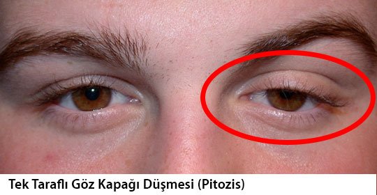
The healthy eyelid covers the colored part of the eye by 1-1.5 mm. If this rate is higher, it is considered as a droopy eyelid. When the droop is unilateral, a difference of more than 2 mm between the two valve heights facilitates the diagnosis of the disease. The most common form of eyelid diseases is drooping of the upper lid, called “ptosis”.
Ptosis may be congenital or may occur later. Congenital ptosis is usually caused by the inability of the muscle that lifts the eyelid to develop properly, while ptosis that occurs in advanced ages is usually caused by the thinning of the muscle that lifts the eyelid and separating from the lid due to old age. In addition, ptosis may occur due to damage to the nerves controlling this muscle or some muscle diseases or injuries. In addition, ptosis may develop after some interventions with speculums or injections to open the eye during other eye surgeries. Transient ptosis may also occur after Botox® injections around the upper eyelid. In addition, injuries, prolonged use of contact lenses, infections, allergic diseases, tumors, some muscle and nerve diseases can cause droopy eyelids.
When ptosis is present, patients try to see by raising their eyebrows or pushing their chin forward and head back. The problem can sometimes even cause the eye to close completely. In this case, patients try to see by lifting their eyelids with their hands.
If droopy eyelids affect vision in children, early surgery is required to prevent amblyopia. In case of droopy eyelids that do not affect vision, surgery is a must in the pre-school period at the latest, and an expert ophthalmologist should be consulted on this issue. Lid droop does not improve with the growth of children. The lack of pre-school treatment negatively affects the child’s personal development.
If double vision is added to the miscarriage, it is necessary to pay attention. Opening of the eyelids than normal is a sign of thyroid gland disease, and closing is a sign of facial paralysis. Double vision with droopy eyelid may also be an indicator of diabetes, some progressive muscle diseases or myasthenia. Those who go to the hospital due to droopy eyelids should be examined in detail. In the examination, old pictures should be requested and it should be noted how long the changes in the eye and its surroundings have been. The patient’s treatment should be planned in the best way by performing the necessary neurological and blood tests and consulting with the relevant departments.
Unexplained drooping is a marker of brain tumor, and subsequent drooping may be caused by some nerve and muscle diseases. The most important ones are brain tumor and vascular diseases; If there has been a drooping of the eyelid without any known reason such as injury, allergy or infection, examinations such as brain and orbital MRI, EMG must be performed and the cause must be found.
If ptosis is present from birth or occurs within the first 10 years of life, it can lead to permanent vision loss. All diseases that impair the quality of vision in the first years of life cause amblyopia, and therefore, an ophthalmologist should be consulted to understand whether congenital or early-onset droopy eyelids impair vision. Lid drooping that occurs in advanced ages, on the other hand, causes vision loss if it covers the pupil, and creates an aesthetic defect if it does not. Permanent vision loss is not expected in these cases.
Since congenital droopy eyelids that are advanced enough to cover the pupil will prevent vision development and cause amblyopia, the timing of treatment is important in these patients and urgent correction of droopy eyelids is required. Mild droopy eyelids, which cause only cosmetic problems without affecting vision, should be corrected just before the child starts school, that is, around the age of 5-6, so that the child feels more comfortable in society and does not adversely affect his psychological development. Ptosis is treated surgically. In mild congenital drooping of the lids and drooping of the lids due to aging, the muscle that lifts the eyelid is shortened and strengthened in the surgery. In severe congenital ptosis due to nerve palsy or with very little muscle function, the eyelid is hung on the forehead muscle of the patient with silicone bands under the skin, so that the patient can open his eyes by raising his eyebrows. Much more beneficial results are obtained if neurosurgeons, neurology and ophthalmologists evaluate those with unilateral droopy eyelids together.